


“
Solid quality control. The numbers on the report match what I received.
A
Adam W.
Verified
Portland OR · Feb 2026
This batch of PEG MGF Peptide has been third party lab tested and verified for quality.
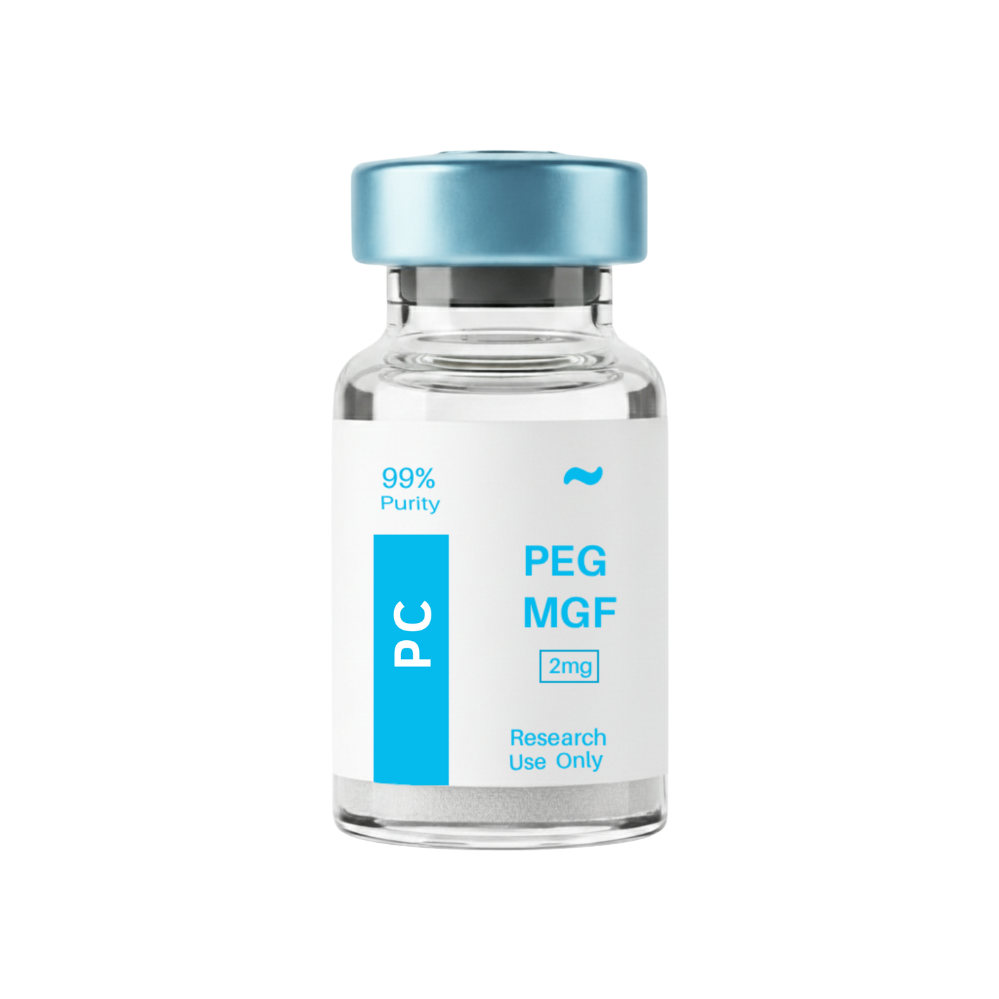
Contents: PEG-MGF
Form: Powder
Purity: 99.3%
View Third-Party Tests from Our Partners:

Couldn't load pickup availability
This product is Shipped & Tested in Canada.
We are committed to delivering fast, reliable, and transparent shipping for all orders. Please review our policy below for details on delivery times, tracking, and what to expect with every purchase.
View Full Shipping Policy


Manufactured in facilities meeting stringent cGMP requirements.
Reliable 3–5 day shipping guaranteed.
Our expert team is ready to help 24/7.


Here you'll find answers to common questions.
Every vial we sell comes from a lab that follows current Good Manufacturing Practices (cGMP). That means each step of production is documented and controlled. Before a batch is released, it’s tested by independent third-party labs for purity, identity, and sterility. Certificates of analysis are available so you can see the exact test results.
Yes. The labs we work with use ISO-certified clean rooms where air quality, equipment, and handling procedures are tightly regulated. Staff are trained to pharmaceutical-grade standards. This ensures the peptides are produced in an environment that minimizes contamination risks.
Peptides in lyophilized (freeze-dried) form are stable at room temperature for transport. Once you receive them, refrigeration is recommended to maintain long-term integrity. We package every order securely to prevent damage and ship promptly, so your vials arrive in optimal condition.
We operate under strict in-house protocols that follow current Good Manufacturing Practices (cGMP). That means our team oversees the entire process from sourcing raw amino acids to the final lyophilized vial. Nothing is outsourced or repackaged. This gives us full control over purity, consistency, and sterility, and it’s why we can stand behind every single vial we ship.
Store them in the refrigerator, away from direct light and heat. If you need to keep them longer, some peptides can be stored frozen. Each vial comes with clear handling instructions so you know the proper conditions for stability.
The strongest proof is transparency. For every peptide, we can provide certificates of analysis, manufacturing documentation, and references to the published scientific research behind it. If you ever have questions, we’ll show you the data rather than ask you to take our word for it.
The difference is transparency. Most sites give you a product name and a price. We provide full batch testing, lab documentation, and direct access to certificates of analysis so you don’t have to guess what you’re getting. When you order from us, you know exactly what’s in the vial, where it was made, and how it was verified.
Solid quality control. The numbers on the report match what I received.
Discreet packaging and quick delivery. Will be ordering again.
The testing transparency is a big deal for me and they deliver on it.
Everything checked out against the certificate. Confident reordering.
Arrived fast and the COA matched the batch number on the vial exactly. Reconstituted clean with no cloudiness.
I appreciate that the HPLC and mass-spec reports are public. Purity was exactly as stated.
Packaging was discreet and well insulated. The lyophilized powder looked intact and sealed properly.
Good quality and quick shipping. Would like a little more handling info on the listing itself.
Third-party testing is the reason I switched. The documentation is thorough and easy to find.
Cold-chain shipping held up. The ice pack was still cold on arrival. Solid all around.
Batch traceability is exactly what I needed for my records. The lot number lines up with the report.
Consistent results across two orders. The included reconstitution solution was a nice touch.
Quality is there. Shipping took five days but tracking was accurate the whole way.
The certificate of analysis is linked right on the site, no chasing support to get it.
Vials sealed properly, labels clear with batch and date. Looks professional.
Customer service answered fast and sent the lab report the moment I asked.